
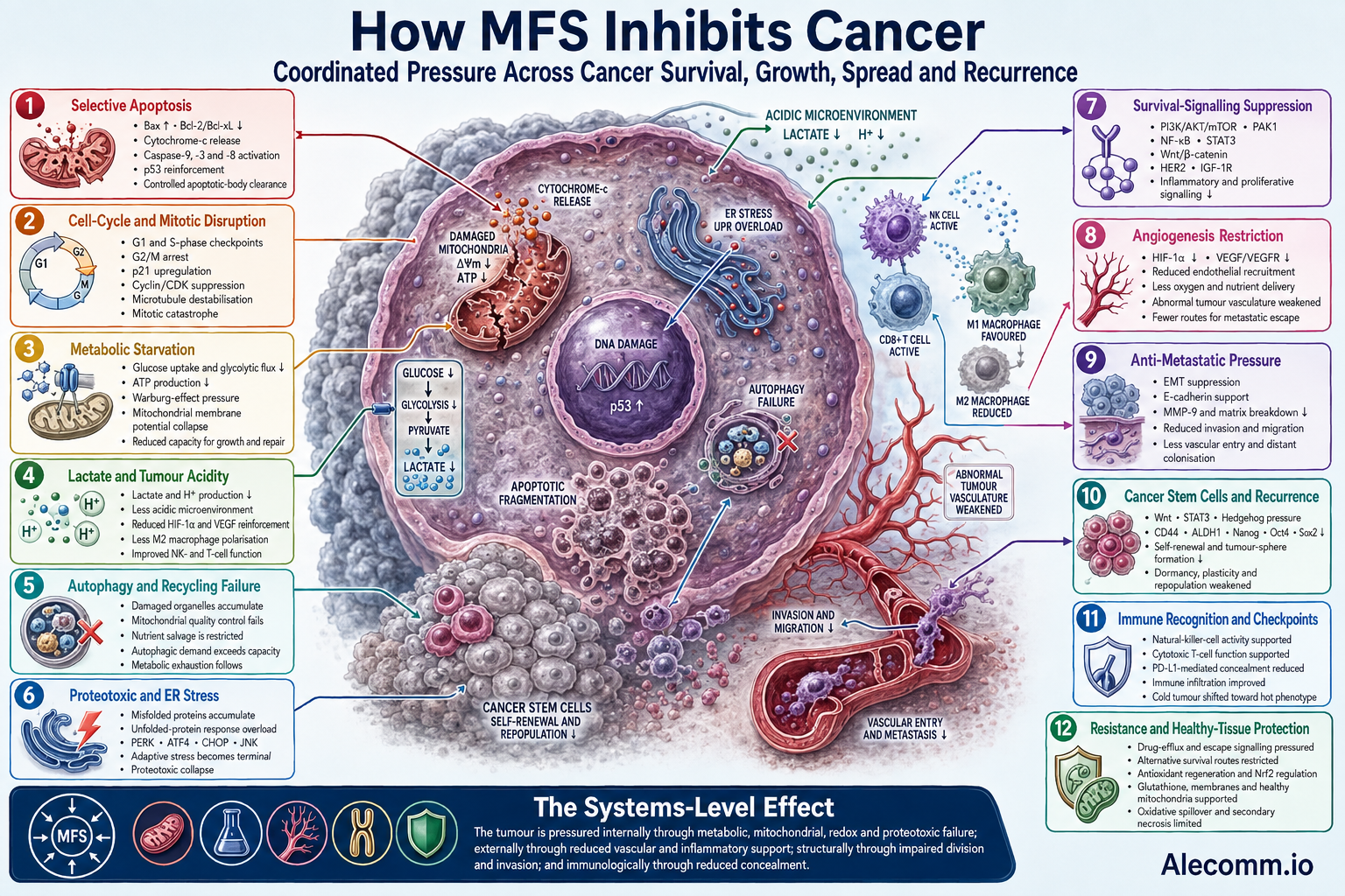
MFS represents a systems-biology approach to cancer support. Rather than relying on single-target cytotoxicity, the formulation works through coordinated, multi-pathway actions that selectively pressure malignant cells while protecting and restoring healthy ones.
The net effect is clean, non-inflammatory programmed cell death in abnormal cells, disruption of tumour growth and survival signalling, metabolic starvation of cancer cells, reduced angiogenesis and metastasis, suppression of cancer stem cells, weakening of treatment resistance, and enhanced immune surveillance.
Cancer survives because it adapts. It changes fuel sources, repairs damage, suppresses immune recognition, recruits new blood vessels, manipulates surrounding tissue, enters dormancy, and preserves stem-like cells capable of rebuilding the disease. The strategic purpose of MFS is to apply pressure across these interconnected survival systems simultaneously.
Core Mechanism: Selective Apoptosis
Cancer cells characteristically display elevated reactive oxygen species, defective mitochondria, genetic instability, abnormal protein production, and overactive survival signalling. MFS exploits these vulnerabilities to restore the cell’s intrinsic ability to recognise irreparable damage and commit to orderly death.
Early Decision Phase: Cell Shrinkage And Chromatin Condensation
The mitochondrial membrane potential collapses, reducing the malignant cell’s ability to generate energy and maintain internal stability.
Pro-apoptotic proteins such as Bax increase, while anti-apoptotic proteins including Bcl-2 and Bcl-xL decline. The balance therefore shifts away from survival and towards mitochondrial membrane permeabilisation.
Cytochrome c is released from damaged mitochondria, contributing to activation of caspase-9 and caspase-3. Death-receptor signalling can also activate caspase-8, connecting external cellular death signals with internal mitochondrial apoptosis.
The tumour-suppressor pathway involving p53 is reinforced, transcription begins shutting down, and chromatin condenses as the cell prepares for controlled dismantling.
Structural Dismantling: Membrane Blebbing
Cytoskeletal collapse follows through actin-filament cleavage and microtubule destabilisation. The plasma membrane detaches from its underlying structural supports, while controlled osmotic and ionic changes produce membrane protrusions known as blebs.
Unlike necrosis, the membrane remains substantially intact during this process. Cellular contents are therefore contained rather than explosively released into surrounding tissue.
Final Clearance: Organelle Disintegration And Packaging
Mitochondria fragment, the endoplasmic reticulum breaks down, nuclear DNA is systematically cleaved, and the dying cell separates into membrane-bound apoptotic bodies.
These fragments display molecular signals that attract macrophages and other phagocytic cells. The abnormal cell can then be removed without provoking the widespread inflammation associated with uncontrolled cellular rupture.
Protective redox buffering helps prevent oxidative spillover and secondary injury in neighbouring healthy tissue.
The result is non-inflammatory apoptosis rather than chaotic necrosis. Healthy cells, possessing more stable mitochondria and balanced redox regulation, are supported rather than indiscriminately pushed towards death.
Multiple Routes To Cellular Death
Apoptosis is the central mechanism, but malignant cells frequently acquire mutations that allow them to evade it. Cancer can disable p53, increase Bcl-2 expression, suppress death receptors, block caspase activation, or activate alternative survival pathways.
MFS therefore applies pressure across several routes of cellular failure:
- Intrinsic apoptosis through mitochondrial membrane collapse, cytochrome-c release, and caspase activation.
- Extrinsic apoptosis through death-receptor signalling and caspase-8 activation.
- Mitotic catastrophe when severely damaged cells attempt to divide but cannot correctly organise or separate their chromosomes.
- Autophagic collapse when cellular recycling can no longer compensate for accumulating mitochondrial, protein, and metabolic damage.
- Irreversible senescence when damaged malignant cells permanently lose their ability to divide.
- Proteotoxic death when abnormal proteins overwhelm the endoplasmic reticulum and cellular quality-control machinery.
Applying pressure across several death pathways reduces the likelihood that blocking one molecular mechanism will be sufficient to preserve the malignant cell.
Disruption Of Cell Division And Cytoskeleton
MFS interferes with the structural machinery required for rapid proliferation. Microtubule destabilisation arrests malignant cells during the G2/M phase, preventing the formation or proper operation of the mitotic spindle.
Without a functioning spindle, duplicated chromosomes cannot be accurately separated between daughter cells. The resulting mitotic failure activates checkpoint responses and can lead to apoptosis, senescence, or mitotic catastrophe.
Additional cell-cycle checkpoints are engaged through:
- Upregulation of p21 and related cell-cycle inhibitors
- Downregulation of cyclins and cyclin-dependent kinases
- G1-phase arrest before DNA replication
- S-phase disruption during DNA synthesis
- G2/M arrest before or during mitosis
This halts mitotic progression in rapidly dividing malignant cells while placing substantially less pressure on healthy quiescent tissue.
Metabolic Interference And The Warburg Effect
Many cancers rely heavily on aerobic glycolysis, known as the Warburg effect, producing energy from glucose even when oxygen is available.
Although glycolysis is less efficient than mitochondrial oxidative phosphorylation, it provides rapidly dividing cells with metabolic intermediates needed to manufacture nucleic acids, proteins, lipids, and cellular membranes.
MFS compounds impair glucose uptake, glycolytic flux, ATP generation, and metabolic signalling within tumour cells. As accessible energy declines, malignant cells experience increasing metabolic stress and reduced capacity to maintain growth, repair damage, operate drug-efflux pumps, or resist programmed death.
At the same time, mitochondrial function within malignant cells is further destabilised through:
- Decreased mitochondrial membrane potential
- Increased reactive oxygen species
- Impaired respiratory-chain activity
- Reduced ATP generation
- Disrupted mitochondrial quality control
- Activation of mitochondrial death signalling
Healthy mitochondria receive support for efficient oxidative phosphorylation, membrane stability, redox regulation, and energy recovery. The resulting metabolic pressure selectively disadvantages malignant cells already operating close to their energetic and oxidative limits.
Tumour Acidity, Lactate, And Metabolic Immune Evasion
The Warburg effect does considerably more than supply energy. It generates large quantities of lactate and hydrogen ions, producing an acidic tumour microenvironment that actively protects malignant cells.
High lactate concentrations suppress cytotoxic T cells and natural-killer cells, reduce interferon-γ production, encourage regulatory T-cell activity, and shift macrophages towards the tumour-supporting M2 phenotype.
Lactate can also reinforce:
- HIF-1α signalling
- VEGF production
- PI3K/AKT/mTOR activity
- JAK/STAT signalling
- Angiogenesis
- Cellular migration and invasion
Lactate also participates in histone lactylation, an epigenetic process through which tumour metabolism can alter gene expression and reinforce tumour-supporting cellular programmes.
By restricting glycolytic efficiency and weakening the pathways that sustain hypoxia, inflammation, and lactate production, MFS attacks both the tumour’s energy supply and its acidic defensive perimeter.
This makes the surrounding environment less favourable to immune suppression, angiogenesis, tissue invasion, and metastatic spread.
Autophagy And The Cancer Cell’s Recycling System
Cancer cells survive hostile conditions by recycling damaged proteins, lipids, and organelles through autophagy.
This internal salvage system supplies nutrients during glucose deprivation, removes damaged mitochondria, reduces toxic protein accumulation, and helps malignant cells withstand hypoxia, oxidative stress, and treatment.
MFS disrupts the balance between protective autophagy and cellular destruction. As mitochondrial damage, oxidative pressure, protein misfolding, and energy depletion accumulate, the tumour cell becomes increasingly dependent upon its recycling machinery.
Eventually, the demand exceeds the cell’s ability to maintain autophagic control. The recycling system can then cease functioning as a survival mechanism and contribute to:
- Autophagic collapse
- Accumulation of damaged mitochondria
- Failure of protein quality control
- Metabolic exhaustion
- Apoptosis
- Irreversible loss of cellular viability
This creates a biochemical trap: the malignant cell becomes increasingly dependent upon its emergency recycling system precisely as its capacity to operate that system is being undermined.
Endoplasmic-Reticulum Stress And Proteotoxic Collapse
Rapidly dividing cancer cells manufacture enormous quantities of proteins. Genetic instability, abnormal chromosome numbers, oxidative stress, and defective metabolism cause many of these proteins to be incorrectly folded.
This places continuous pressure on the endoplasmic reticulum and unfolded-protein response.
Cancer cells initially use this response to pause protein production, remove defective proteins, conserve energy, and avoid death. When the damage becomes sustained, however, the same protective system changes direction.
Signalling through PERK, ATF4, CHOP, JNK, and mitochondrial death pathways converts an adaptive stress response into a terminal one.
MFS intensifies this vulnerability by combining:
- Mitochondrial disruption
- Redox pressure
- Impaired energy production
- Abnormal protein accumulation
- Autophagic overload
- Suppression of survival signalling
Once the malignant cell can no longer restore protein quality control, it crosses from temporary stress into proteotoxic collapse and programmed death.
Suppression Of Key Survival And Growth Pathways
Multiple overlapping signalling axes sustain tumour growth, proliferation, invasion, immune evasion, and treatment resistance. MFS places pressure on several of these systems simultaneously.
PI3K/AKT/mTOR And PAK1
The PI3K/AKT/mTOR pathway regulates nutrient sensing, glucose metabolism, protein synthesis, cellular growth, autophagy, and resistance to apoptosis.
PAK1 interacts with cytoskeletal organisation, cellular movement, proliferation, and survival signalling. Attenuation of these pathways reduces the malignant cell’s ability to grow, migrate, adapt metabolically, and resist programmed death.
NF-κB
The NF-κB pathway sustains chronic inflammation and activates genes involved in cellular survival, invasion, angiogenesis, and treatment resistance.
Its suppression reduces tumour-supporting mediators including IL-6, TNF-α, COX-2, and MMP-9, weakening the inflammatory environment on which many cancers depend.
STAT3
Persistent STAT3 activation promotes proliferation, cancer stemness, angiogenesis, immune suppression, and resistance to apoptosis.
Suppressing STAT3 reduces the tumour’s ability to maintain stem-like cells, recruit vascular support, and prevent immune-mediated clearance.
Wnt/β-Catenin
Abnormal Wnt/β-catenin signalling supports cellular proliferation, stemness, epithelial-to-mesenchymal transition, invasion, and metastatic development.
Restraining this pathway reduces transcription of genes that allow malignant cells to reproduce, remain undifferentiated, and acquire invasive characteristics.
Growth-Factor Receptors
Phosphorylation and downstream signalling through HER2 and IGF-1R are inhibited in relevant experimental models.
These receptors communicate with PI3K/AKT/mTOR, MAPK, NF-κB, and hormone-receptor pathways. Their suppression therefore removes several overlapping signals for proliferation and survival.
Angiogenic Drivers
Reduced VEGF expression, VEGFR signalling, and HIF-1α activity limits the formation of new blood vessels required to supply expanding tumours with oxygen and nutrients.
These coordinated actions starve tumours of growth signals, inflammatory support, metabolic flexibility, and vascular supply.
Epigenetic Reprogramming
Cancer is driven not only by mutations but also by abnormal control over which genes are switched on or silenced.
Altered DNA methylation, histone modification, chromatin structure, and microRNA activity can silence tumour-suppressor genes while maintaining inflammatory, proliferative, stem-like, and metastatic programmes.
MFS influences signalling connected with:
- DNA methyltransferases
- Histone deacetylases
- Sirtuins
- Nrf2
- p53
- NF-κB
- Wnt/β-catenin
- Tumour-regulating microRNAs
These effects can shift transcription away from uncontrolled proliferation, stemness, angiogenesis, inflammatory survival, and metastatic behaviour.
This adds another level to the formulation’s strategic logic. MFS does not only place pressure on the proteins produced by malignant genes. It also affects the regulatory machinery determining which genes the cancer is permitted to express.
Anti-Angiogenesis And Vascular Starvation
A tumour cannot continue expanding without constructing its own blood supply. Hypoxic cancer cells release HIF-1α and VEGF, stimulating nearby endothelial cells to develop abnormal blood vessels.
These vessels supply nutrients but are structurally disorganised and permeable, contributing to tumour inflammation, hypoxia, and the movement of malignant cells into circulation.
By lowering VEGF, VEGFR signalling, and HIF-1α, MFS reduces the biological signals required for tumour vascularisation.
Reduced vascular support creates additional metabolic pressure by limiting:
- Oxygen delivery
- Glucose availability
- Amino-acid supply
- Removal of metabolic waste
- Access to routes of metastatic escape
This vascular restriction works alongside glycolytic interference and mitochondrial disruption to intensify tumour energy stress.
Anti-Metastatic Activity And Microenvironment Modulation
Metastasis requires cancer cells to detach from the primary tumour, invade surrounding tissue, enter blood or lymphatic vessels, survive circulation, exit into distant organs, and establish new colonies.
MFS places pressure on several stages of this process.
Epithelial-to-mesenchymal transition is inhibited, reducing the transformation through which stationary epithelial cells acquire mobile and invasive properties.
Cell-adhesion molecules such as E-cadherin are supported, helping cells retain normal structural attachment. Matrix metalloproteinases, including MMP-9, are reduced, limiting the degradation of extracellular barriers surrounding the tumour.
The tumour microenvironment is shifted away from a pro-tumour state characterised by:
- Chronic inflammation
- Hypoxia
- Lactate accumulation
- M2 macrophage dominance
- Immune suppression
- Extracellular-matrix breakdown
- Abnormal vascularisation
The combined effect is reduced capacity for invasion, vascular entry, metastatic survival, and colonisation of distant tissue.
Cancer Stem Cells, Dormancy, And Recurrence
Destroying the visible tumour is not always enough. A small population of cancer stem cells can survive treatment, remain dormant, and later regenerate the disease.
These cells possess enhanced DNA repair, antioxidant capacity, drug-efflux systems, metabolic flexibility, and the ability to shift between dormant and rapidly proliferating states.
MFS applies pressure to signalling networks that maintain this stem-like population, including:
- Wnt/β-catenin
- NF-κB
- STAT3
- PI3K/AKT/mTOR
- Hedgehog signalling
- Epithelial-to-mesenchymal transition pathways
Suppression of these networks reduces self-renewal, tumour-sphere formation, cellular plasticity, and expression of stemness-associated markers such as CD44, ALDH1, Nanog, Oct4, and Sox2.
This is strategically important because cancer stem cells are strongly associated with recurrence, metastasis, treatment resistance, and repopulation of the tumour after apparent remission.
The objective is therefore not merely to reduce tumour bulk, but to weaken the cellular reservoir capable of rebuilding it.
Immune Modulation And Checkpoint Support
Tumours survive partly by concealing themselves from the immune system. They can suppress antigen presentation, exhaust T cells, recruit regulatory immune cells, and express checkpoint molecules that effectively instruct immune cells not to attack.
MFS supports natural-killer-cell and T-cell activity, increases immune-cell infiltration into tumours, and can reduce immune-evasion molecules such as PD-L1.
A poorly infiltrated immunologically “cold” tumour may therefore be shifted towards a more active “hot” phenotype with greater potential for immune recognition.
MFS also reduces chronic inflammatory cytokines that paradoxically promote tumour progression while weakening effective anti-tumour immunity.
Its immune actions include:
- Supporting natural-killer-cell recognition of abnormal cells
- Strengthening cytotoxic T-cell function
- Reducing tumour-associated immune suppression
- Limiting M2 macrophage polarisation
- Reducing inflammatory survival signals
- Improving the environment for immune-cell infiltration
The purpose is not indiscriminate immune stimulation, but the restoration of more effective immune surveillance, recognition, and clearance of abnormal cells.
Treatment Resistance And Re-Sensitisation
Treatment resistance develops through overlapping adaptations, including:
- Increased drug export
- Stronger DNA repair
- Enhanced antioxidant buffering
- Altered glucose and mitochondrial metabolism
- Cancer-stem-cell survival
- Hypoxia
- Apoptosis resistance
- Autophagic recycling
- Activation of alternative growth pathways
MFS places concurrent pressure on many of these resistance systems.
Suppression of Bcl-2, NF-κB, STAT3, PI3K/AKT/mTOR, Wnt/β-catenin, HIF-1α, VEGF, and drug-efflux signalling reduces the number of escape routes available to the tumour.
This multi-node strategy is important because cancer rarely relies upon a single pathway. When one pathway is blocked, malignant cells frequently reroute signalling through another.
Simultaneous disruption makes that biological rerouting progressively more difficult and can increase tumour susceptibility to additional therapeutic pressures.
Hormone-Dependent Tumour Signalling
Certain breast, ovarian, endometrial, and prostate cancers remain dependent upon hormone-driven growth.
MFS contains mechanisms relevant to oestrogen metabolism, aromatase activity, androgen-receptor signalling, inflammatory hormone synthesis, and growth-factor cross-talk.
Hormone receptors do not operate independently. They communicate with:
- HER2
- IGF-1R
- PI3K/AKT/mTOR
- MAPK
- NF-κB
Pressure applied across these interconnected systems reduces the ability of hormone-dependent tumours to bypass receptor suppression through alternative survival signalling.
Redox Regulation, Detoxification, And Tissue Protection
Cancer cells already operate under elevated oxidative pressure. Their rapid metabolism, abnormal mitochondria, protein production, and genetic instability generate large quantities of reactive oxygen species.
MFS pushes this internal stress towards cytotoxic levels within malignant cells while supporting redox regulation in healthy tissue.
This apparently contrasting activity is possible because malignant and healthy cells begin from very different biochemical positions. Cancer cells frequently exist close to their maximum tolerable oxidative threshold, whereas healthy cells retain more effective mitochondrial regulation, antioxidant recycling, and stress-response control.
MFS supports:
- Antioxidant regeneration
- Nrf2-pathway regulation
- Glutathione metabolism
- Membrane stabilisation
- Mitochondrial integrity
- Detoxification pathways
- DNA repair and methylation
- Healthy-cell energy production
These actions help prevent oxidative spillover, secondary inflammation, and necrotic injury in surrounding tissue while maintaining genomic integrity during periods of cellular stress.
Cancer Types In Which These Mechanisms Have Been Observed
Preclinical and mechanistic evidence associated with components and pathways represented within MFS spans a wide range of malignancies:
- Breast cancers, including triple-negative and HER2-overexpressing subtypes
- Colorectal and other gastrointestinal cancers
- Prostate cancer
- Lung cancers, including adenocarcinoma and non-small-cell lung cancer
- Pancreatic cancer
- Renal-cell carcinoma
- Glioblastoma and other brain tumours
- Hepatocellular carcinoma
- Leukaemias and lymphomas
- Ovarian cancer
- Cholangiocarcinoma
- Gastric cancer
- Melanoma
- Other solid tumours displaying comparable metabolic and signalling abnormalities
Across these cancers, the relevant targets include mitochondrial dysfunction, glycolytic addiction, constitutive survival signalling, angiogenesis, stemness, immune evasion, metastatic plasticity, and chronic inflammation.
The strategy does not depend upon a single molecular lesion shared by every tumour. It addresses biological systems repeatedly exploited across different cancer types.
The Wider Systems Effect
The anti-cancer rationale becomes clearest when MFS is understood as a coordinated biological network rather than a collection of isolated agents.
- Metabolic pressure deprives malignant cells of readily available energy.
- Mitochondrial disruption restricts their ability to compensate through respiration.
- Redox pressure pushes already stressed cancer cells beyond their survival threshold.
- Cell-cycle arrest prevents damaged cells from reproducing.
- Mitotic disruption makes successful chromosome separation increasingly difficult.
- Proteotoxic stress overwhelms protein quality-control systems.
- Autophagic disruption undermines the cancer cell’s emergency recycling machinery.
- Stemness suppression attacks the population responsible for recurrence and tumour repopulation.
- Anti-angiogenic activity restricts oxygen and nutrient delivery.
- Microenvironment modulation weakens acidic, inflammatory, and metabolic protection.
- Anti-metastatic activity reduces tissue invasion and distant colonisation.
- Immune support improves recognition and clearance of abnormal cells.
- Healthy-tissue support protects mitochondrial function, membrane integrity, redox balance, detoxification, and recovery capacity outside the tumour.
The result is a form of systems-level containment. The tumour is pressured internally through metabolic, mitochondrial, proteotoxic, and redox failure; externally through reduced vascular and inflammatory support; structurally through impaired division and invasion; and immunologically through reduced concealment.
Overall Strategic Logic
MFS does not rely upon indiscriminate cellular destruction. It restores pressure towards programmed death in cells carrying irreparable damage, starves malignant cells of energy and growth signals, restricts their blood supply, undermines their ability to recycle damaged components, reduces metastatic potential, suppresses stem-like populations, and strengthens immune surveillance.
Its significance lies in the convergence of these mechanisms. Cancer cells can often survive the inhibition of one pathway by activating another. It becomes considerably harder for them to escape when energy production, mitochondrial stability, cell division, angiogenesis, inflammatory signalling, cellular recycling, stemness, immune evasion, and metastatic behaviour are pressured at the same time.
Cancer survives by adapting. It changes fuel sources, reroutes signalling, repairs damage, suppresses immunity, recruits blood vessels, enters dormancy, and preserves cells capable of rebuilding the disease.
MFS confronts that adaptability with coordinated pressure across the same interconnected systems. Its central strength is not one isolated anti-cancer action, but the simultaneous disruption of the biological network that allows malignant cells to survive, spread, resist treatment, and return.
This multi-layered, systems-level inhibition of cancer initiation, progression, adaptation, and survival pathways constitutes the core anti-cancer rationale of MFS.
Sources And References
- Jiang, X., et al. (2024). The regulation and function of Nrf2 signalling in ferroptosis-activated cancer therapy. PubMed
- Pouysségur, J., et al. (2022). The Warburg effect controls tumour growth, immunity, and microenvironmental acidosis. PubMed
- Yu, X., et al. (2024). Histone lactylation: From tumour lactate metabolism to gene regulation within the tumour microenvironment. PubMed
Leave a Reply